Gallery
adipocyte images: illustrations and micrographs. All credits and licenses listed.
Micrographs
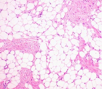
White adipose tissue, H&E (lipoma section). Mature unilocular adipocytes with the classic signet-ring appearance: large empty spaces after lipid extraction, thin cytoplasm rims, and peripheral haematoxylin-stained nuclei. Fibrous capsule and septal stroma visible.
Hyeree Kim, Sang Hyun Cho, Jeong Deuk Lee, Hei Sung Kim, Wikimedia Commons · CC BY 4.0 · source
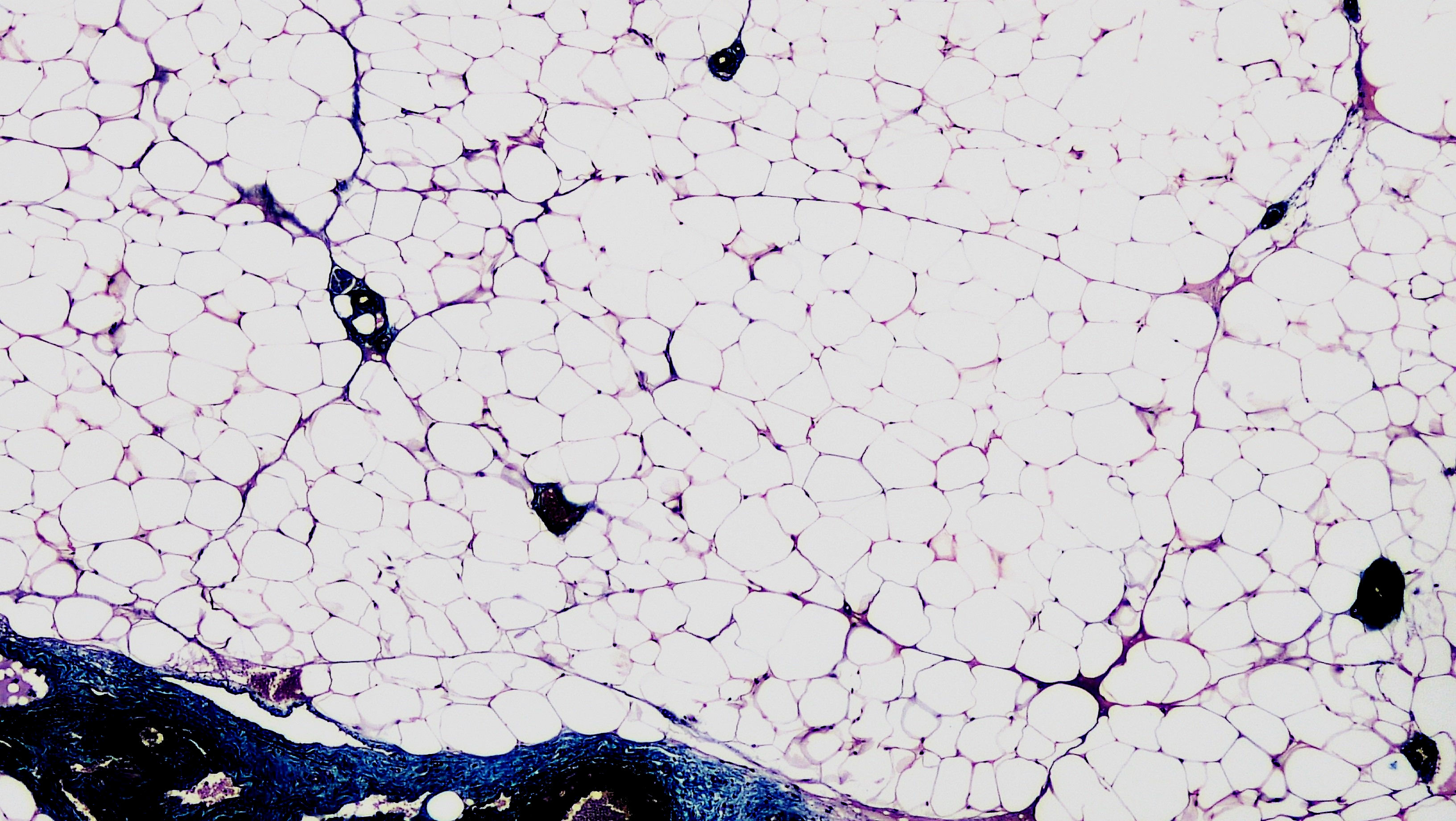
White adipose tissue, histological section. Unilocular adipocytes in connective tissue context, showing the lobular architecture of adipose tissue and the relationship between fat cells and their supporting stroma.
Berkshire Community College Bioscience Image Library, Wikimedia Commons · CC0 · source
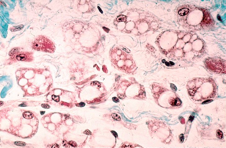
Brown adipose tissue, histological section. Multilocular adipocytes with smaller lipid droplets and cytoplasm-rich cells, distinct from the unilocular white adipocyte. The denser, more granular cytoplasm reflects the high mitochondrial content that drives UCP1-mediated thermogenesis.
Wikimedia Commons · public domain · source
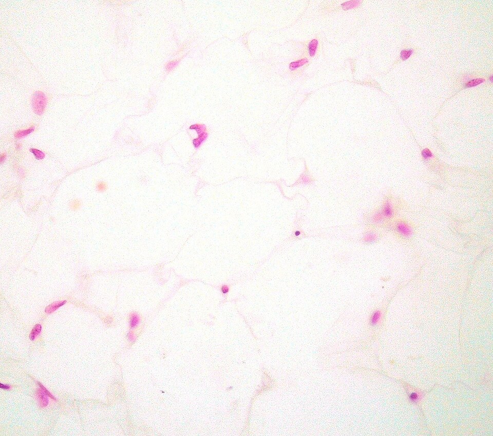
White adipose tissue, H&E (inferred). Adipocytes with lipid voids at the centre and peripheral pink nuclei pushed to the cell rim. Ghost-cell morphology is the expected artifact of paraffin processing: lipid dissolves, leaving a clear sphere outlined by a thin cytoplasmic rim and a flattened crescent nucleus.
Echinaceapallida, Wikimedia Commons · CC BY-SA 4.0 · source
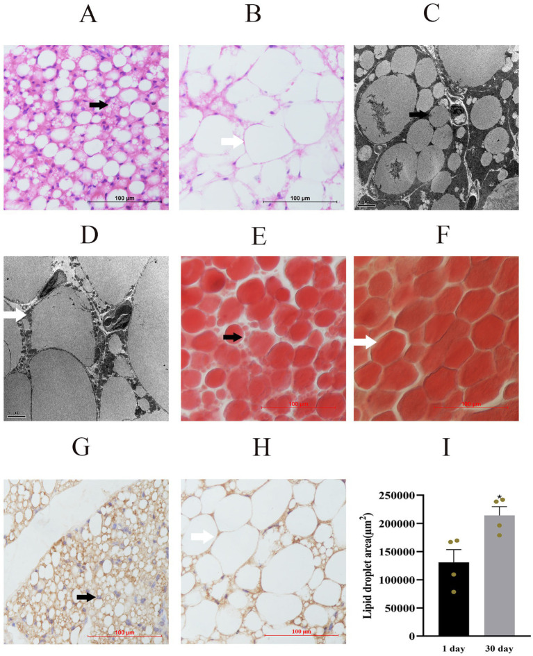
Perirenal adipose tissue in yak calves (H&E, panels A-B; SEM, Oil Red O and UCP1 IHC, panels C-I). Postnatal day 1 and day 30 sections show multilocular brown adipocytes (black arrows) with smaller lipid droplets and mitochondria-rich cytoplasm, contrasting with the large unilocular white adipocytes in the surrounding depot.
Bai, Fan, Hao et al. 2026, Frontiers in Veterinary Science · CC BY 4.0 · source
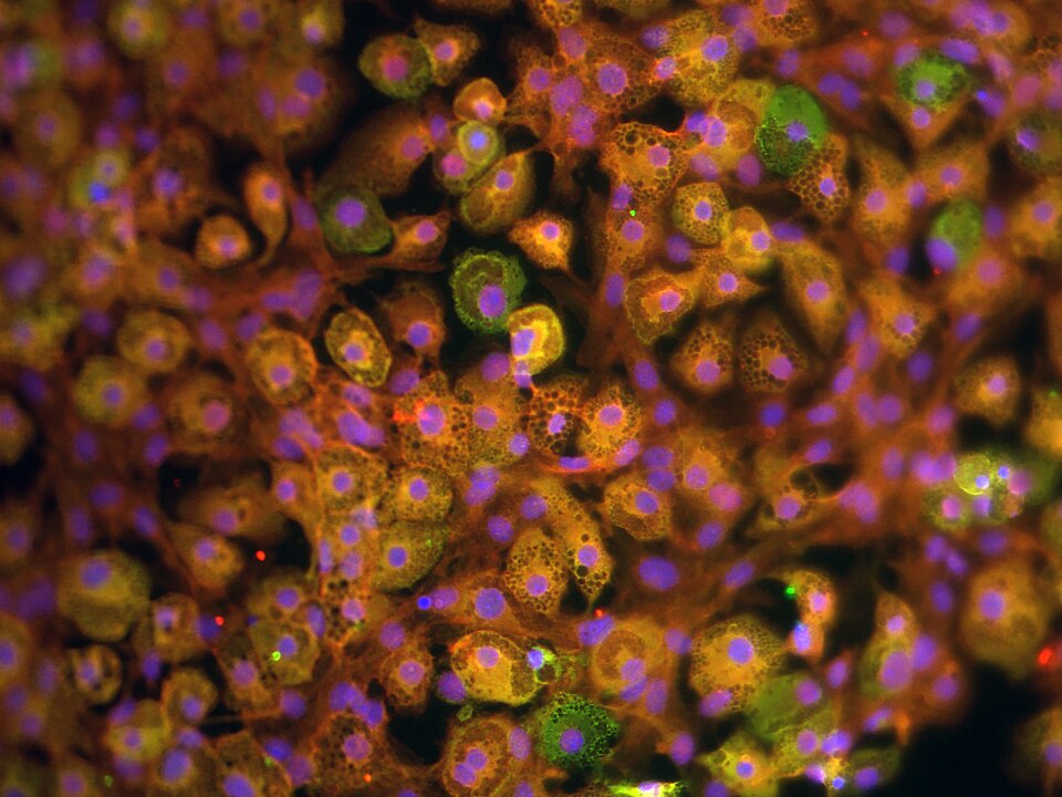
Differentiated adipocytes (PA6 cell line), light microscopy. Round lipid-laden cells with cytoplasm compressed to a thin rim around the central lipid droplet, demonstrating the characteristic unilocular morphology of the white adipocyte.
M. Oktar Guloglu, Wikimedia Commons · CC BY-SA 4.0 · source
Illustrations
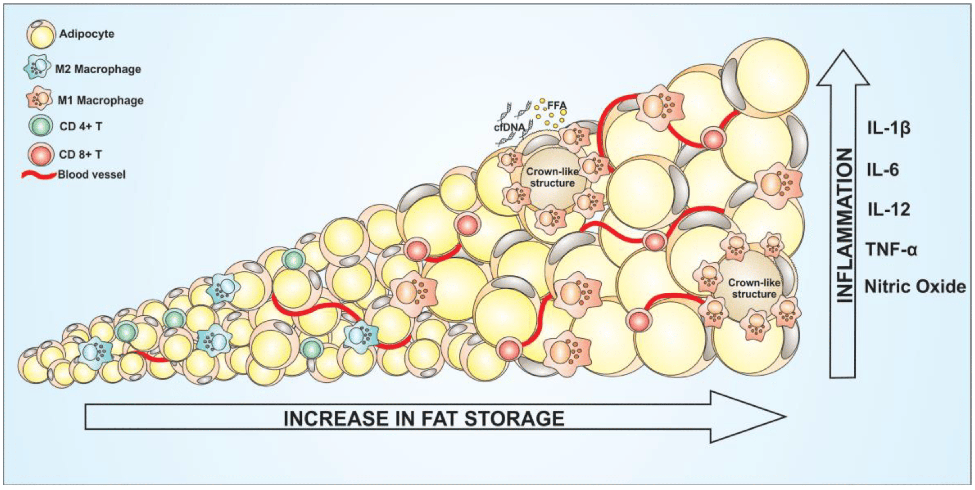
Adipose tissue inflammation in obesity: when an adipocyte hypertrophies beyond its capacity, it undergoes stress and death. Macrophages are recruited and form a crown-like structure around the dead cell, secreting TNF-alpha and IL-6. This diagram shows the lipid-overflow-to-inflammation sequence that links adipocyte hypertrophy to systemic insulin resistance.
Correa et al., Wikimedia Commons · CC BY 4.0 · source
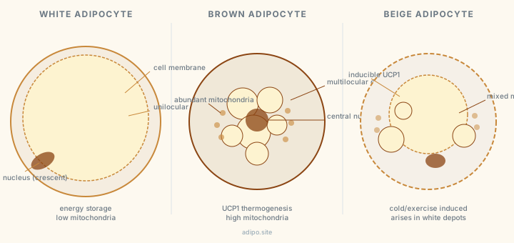
White, brown, and beige adipocytes: structural comparison. White holds a single unilocular lipid droplet with a crescent nucleus pushed to the rim. Brown has multilocular droplets and abundant mitochondria. Beige arises in white depots under cold or adrenergic stimulation and expresses UCP1.
adipo.site · CC0
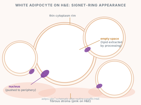
White adipocyte on H&E: the signet-ring appearance. Lipid is extracted by processing solvents, leaving a large empty space with a thin cytoplasm rim and a peripheral crescent nucleus stained purple with haematoxylin. The empty space is not pathology; it is expected paraffin processing artifact.
adipo.site · CC0
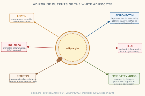
Adipokine outputs of the white adipocyte: leptin, adiponectin, TNF-alpha, IL-6, resistin, and free fatty acids. Each arrow shows the downstream effect: appetite suppression, insulin sensitization, inflammation, IRS-1 serine phosphorylation, and lipotoxicity via ectopic fat deposition.
adipo.site · CC0