adipo.site

Everything known about the adipocyte, past and present.

Building the model…
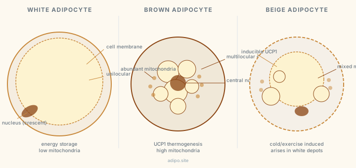
Living model of the adipocyte: the cell. Zoom between the stages. Morphology after Frayn KN, Karpe F, Fielding BA et al., Physiol Rev 2003; Lodhi IJ and Semenkovich CF, Cell Metab 2014; Rutkowski JM, Stern JH, Scherer PE, J Cell Biol 2015.

Adipo. I hold everything known about the adipocyte. What do you need?

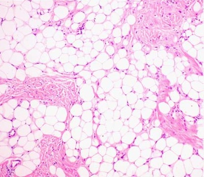
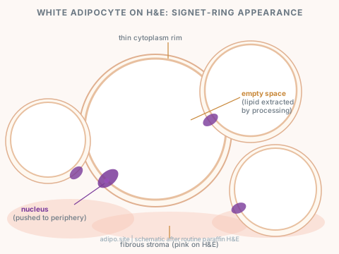
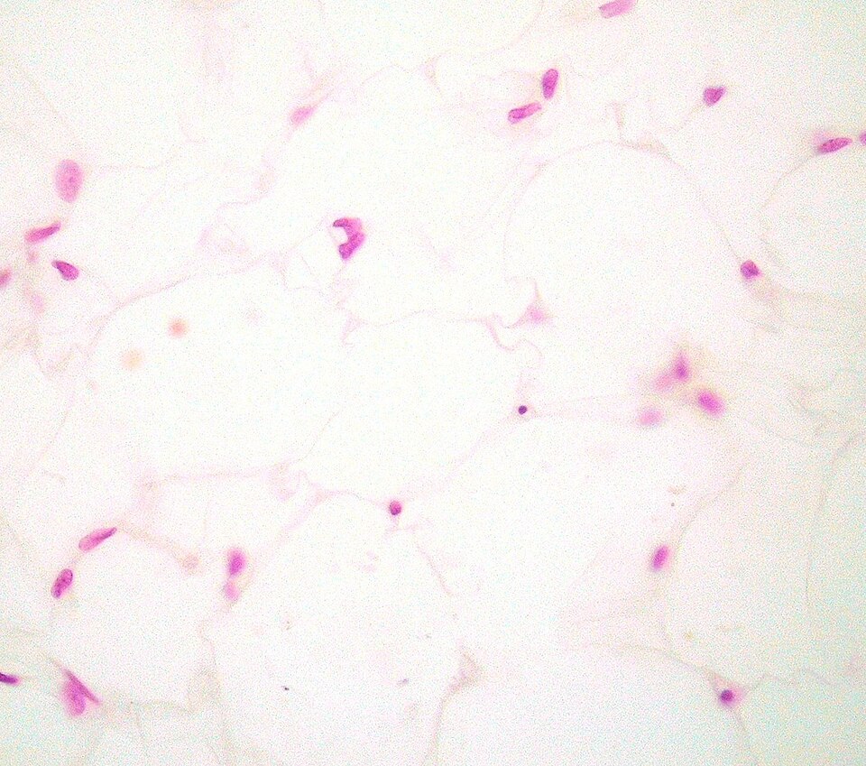
The adipocyte is a round cell built for lipid storage. A mature white adipocyte holds a single unilocular lipid droplet that fills up to 95 percent of its volume, pushing the nucleus and organelles into a thin rim at the plasma membrane. On H&E this produces the signet-ring appearance. The empty space is not pathology; it is what the cell looks like when you dissolve the fat out during processing.

The adipocyte is also an endocrine cell. It secretes leptin, adiponectin, and more than five hundred other signaling molecules that regulate appetite, insulin sensitivity, and systemic inflammation. The fat cell was declared passive until 1994. It was not passive before that either; no one had looked. The master transcriptional regulator of adipocyte differentiation is PPARgamma; forced expression in fibroblasts is sufficient to convert them to adipocytes (Tontonoz et al., 1994).

2026-09-09 · Frontiers in Endocrinology
Editorial: Brown and beige adipocytes, molecular mechanisms to therapeutic potentials

Editorial framing beige and brown adipocyte plasticity as a live therapeutic target for metabolic disease, not just a curiosity.

2026-09-09 · PubMed
Disease-associated adipose browning: current evidence and perspectives

Review of adipose browning tied to disease states, not only cold exposure or exercise.

2026-09-09 · PMC
Adipose Tissue Heterogeneity: Depot-Specific Location and Functional Specialization in Obesity-Related Disease

Depot location (visceral vs subcutaneous vs others) changes adipocyte function and disease risk, not just fat mass.

2026-09-09 · Cell Metabolism
Single-nucleus RNA sequencing identifies mesothelial cells and adipocyte progenitors as strongest correlates of metabolic disease severity

snRNA-seq of subcutaneous and visceral depots from human subjects with varying metabolic disease severity; progenitor cell populations outperform mature adipocyte counts as disease markers.

2026-09-09 · Cell Metabolism
Two distinct beige adipocyte subpopulations identified by snRNA-seq: UCP1-beige and futile-cycle adipocytes

The thermogenic compartment of white adipose tissue is not uniform; UCP1-expressing and futile-cycling subtypes have distinct gene programs and likely distinct therapeutic relevance.

2026-09-09 · Frontiers in Cell and Developmental Biology
Adipose depot traits outperform BMI in predicting cardiometabolic risk

Cellular composition, inflammatory status, and developmental origin of a depot predict disease better than body mass index alone across multiple conditions.

2026-09-10 · Nature Communications
Single-nucleus analysis of human white adipose tissue reveals adipocyte subsets with distinct metabolic profiles

Efthymiou V et al. 2026. snRNA-seq of human WAT identifies adipocyte subpopulations; a low-maturation subset linked to TSHZ3-mediated transcriptional suppression is associated with metabolic disease severity.

2026-09-10 · Biochemical Society Transactions
Understanding adipocyte heterogeneity across species, depot, and disease

Kitto ES et al. 2026. snRNA-seq and spatial transcriptomics reveal previously unappreciated adipocyte subpopulations across depots and disease states; cross-species comparisons clarify conserved vs. human-specific subsets.

2026-09-10 · Molecular Metabolism
Single cell transcriptomics of human weight loss links adipocyte NPY1R to control of lipolysis

Grothen JER et al. 2026. NPY receptor 1 (NPY1R) identified as a cell-autonomous brake on adipocyte lipolysis; expression is inversely correlated with BMI and rises after weight loss, implicating neuropeptide Y signaling in direct lipolysis control.

2026-09-10 · Physiology
Adipose Progenitor Cells in Thermogenesis and Metabolic Regulation

Sahin C et al. 2026. Heterogeneous adipose progenitor subsets are characterized; specific subpopulations are required for thermogenic adipocyte generation and healthy remodeling, with implications for browning strategies.

2026-09-09 · Nature
Positional cloning of the mouse obese gene and its human homologue

Zhang et al. 1994. Leptin identified as the ob gene product, a 16-kDa adipocyte-secreted hormone that acts centrally via the hypothalamus to suppress appetite and regulate energy expenditure.

2026-09-09 · Journal of Biological Chemistry
A novel serum protein similar to C1q, produced exclusively in adipocytes

Scherer et al. 1995. Adiponectin (Acrp30) discovered as a second major adipokine, induced 100-fold during adipocyte differentiation, now known to improve insulin sensitivity via AdipoR1/R2.

2026-09-10 · PMC
Adipocyte browning: mechanisms and anti-obesity pharmacology

Reviews white-to-beige adipocyte transdifferentiation mechanisms and current pharmacological strategies including beta-3 agonists, thyroid hormone analogs, and FGF21 mimetics targeting browning for obesity treatment.

2026-09-09 · New England Journal of Medicine
Semaglutide 2.4 mg for weight management in adults with overweight or obesity (STEP 1 trial)

Wilding et al. 2021. Semaglutide 2.4 mg weekly produced 14.9% mean weight loss vs 2.4% placebo over 68 weeks. First major trial establishing GLP-1RA as primary obesity pharmacotherapy.

2026-09-09 · New England Journal of Medicine
Tirzepatide versus semaglutide for weight loss in obesity (SURMOUNT-5)

Aronne LJ et al. 2025. Tirzepatide (GIP+GLP-1 dual agonist) produced superior weight loss to semaglutide in head-to-head trial; 47% of tirzepatide patients achieved 25% or more weight loss.

2026-09-09 · Cell
PPARgamma as the target of thiazolidinediones: rosiglitazone and pioglitazone bind and activate the receptor

Tontonoz et al. 1994 (extended). TZDs work because PPARgamma is the master adipogenic regulator; activating it redirects lipid to adipose depots and improves systemic insulin sensitivity.

2026-09-09 · Diabetes
Adiponectin levels rise with thiazolidinedione treatment via PPARgamma activation

Maeda et al. 2001. TZDs increase adiponectin secretion from adipocytes as a primary mechanism of their insulin-sensitizing effect, independent of weight loss.

2026-09-09 · Journal of Clinical Investigation
Metreleptin replacement in lipodystrophy: metabolic effects and clinical outcomes

Brown et al. 2012. Metreleptin (recombinant leptin) restores insulin sensitivity and corrects hypertriglyceridemia in leptin-deficient lipodystrophy; the clearest proof of leptin's metabolic role in humans.

2026-09-10 · Physiological Reports
Adipocyte-specific FFA2 deletion leads to increased adipose inflammation and altered intestinal lipid handling

Nnyamah C et al. 2026. Free Fatty Acid Receptor 2 (FFA2/FFAR2) on adipocytes suppresses adipose tissue inflammation; mice lacking adipocyte FFA2 show worsened inflammatory phenotype and disrupted intestinal lipid absorption.

2026-09-10 · Frontiers in Physiology
Impaired adipogenesis drives insulin resistance in non-obese individuals

Defective subcutaneous adipogenesis produces insulin resistance independently of excess adipose mass, expanding the adipocyte dysfunction framework beyond obesity to lean insulin-resistant individuals.

2026-09-10 · PMC
Adipocyte size, overweight, and insulin resistance in T2DM: systematic review

Systematic review of 113 human and 29 animal studies: adipocyte hypertrophy is consistently associated with insulin resistance independent of total adiposity; weight loss reduces cell size and improves insulin sensitivity in parallel.

2026-09-09 · Journal of Clinical Investigation
Obesity is associated with macrophage accumulation in adipose tissue

Weisberg et al. 2003. Up to 40% of obese adipose cells are macrophages. They account for nearly all TNF-alpha in fat; their number tracks adipocyte size and BMI.

2026-09-09 · Journal of Clinical Investigation
Chronic adipose inflammation as a cause of obesity-related insulin resistance

Xu et al. 2003. Macrophage-specific and inflammation genes are upregulated in obese white adipose tissue in mice and humans, proposed as a direct cause of insulin resistance.

2026-09-09 · Arteriosclerosis
Portal adipose tissue as a generator of cardiovascular risk factors

Bjorntorp 1990. The portal hypothesis: visceral fat drains FFA directly to the liver, driving hepatic insulin resistance. Central obesity as a cardiometabolic risk factor explained at the level of anatomy and physiology.

2026-09-10 · Cells
Exploring adipose tissue complexity through omics: implications for health and disease

Sebaa R et al. 2026. Multi-omics review: genomics, transcriptomics, proteomics, and metabolomics of adipose tissue; catalogs how each layer contributes to understanding adipocyte heterogeneity and disease association.

2026-09-09 · Nature
Positional cloning of the mouse obese gene and its human homologue

Zhang et al. 1994. The paper that made adipose tissue an endocrine organ. Leptin identified; field transformed.

2026-09-09 · Cell
Stimulation of adipogenesis in fibroblasts by PPARgamma2

Tontonoz, Hu, Spiegelman 1994. PPARgamma established as master regulator of adipocyte differentiation.

2026-09-09 · FEBS Letters
Bioenergetics of brown adipose tissue mitochondria

Nicholls 1976. UCP1 mechanism. Founding paper for the entire brown adipocyte thermogenesis field.

2026-09-09 · Journal of Biological Chemistry
A novel serum protein similar to C1q, produced exclusively in adipocytes

Scherer et al. 1995. Adiponectin discovered.

2026-09-09 · Science
Adipose expression of TNF-alpha: direct role in obesity-linked insulin resistance

Hotamisligil, Shargill, Spiegelman 1993. Fat as an inflammatory organ.

Substances tested 19

All substances →
Insulin peptide hormone
acts on insulin receptor, then Akt and phosphodiesterase 3B (PDE3B)
Anti-lipolytic. Insulin activates PDE3B in adipocytes through Akt, lowering cAMP and shutting down the PKA-driven lipolytic cascade. Insulin-induced phosphorylation and activation of PDE3B in 3T3-L1 adipocytes was blocked by dominant-negative Akt; the S273A mutant was neither phosphorylated by insulin nor activated in adipocytes, placing Ser273 as the Akt site.
dose: not stated in the abstract  · mouse 3T3-L1 adipocytes  · dominant-negative Akt, site-directed mutagenesis, in vitro kinase and PDE activity assays  · Kitamura T et al. 1999, Mol Cell Biol  · source
Insulin peptide hormone
acts on insulin receptor, then GLUT4 glucose transporter
Stimulates glucose transport by translocating transporters from an intracellular pool to the plasma membrane rather than by activating transporters already at the surface. The founding observation for insulin-regulated GLUT4 trafficking in the fat cell.
dose: not stated in the abstract (the PubMed record for this 1980 paper carries no abstract; title, authors and journal verified)  · rat, isolated adipose cells  · subcellular fractionation and glucose transport measurement  · Cushman SW and Wardzala LJ 1980, J Biol Chem  · source
Mirabegron selective beta-3 adrenergic agonist
acts on ADRB3 (beta-3 adrenergic receptor)
Activates human brown adipose tissue. A single oral dose raised BAT metabolic activity on 18F-FDG PET/CT in all twelve subjects (p = 0.001) and increased resting metabolic rate by 203 plus or minus 40 kcal per day, a 13 percent rise (p = 0.001). BAT activity significantly predicted the change in resting metabolic rate.
dose: 200 mg oral, single dose  · human, 12 healthy men  · 18F-FDG PET/CT with indirect calorimetry, placebo controlled  · Cypess AM et al. 2015, Cell Metab  · source
Rosiglitazone (BRL49653) thiazolidinedione, antidiabetic
acts on PPARgamma (nuclear receptor)
Thiazolidinediones are potent and selective activators of PPARgamma. BRL49653 binds PPARgamma with a dissociation constant near 40 nM. Treating pluripotent C3H10T1/2 stem cells with BRL49653 drove efficient differentiation into adipocytes, linking the antidiabetic drug class directly to the adipogenic master regulator.
dose: Kd approximately 40 nM (binding affinity stated in the abstract)  · mouse C3H10T1/2 pluripotent stem cells  · receptor binding and transactivation assays with differentiation readout  · Lehmann JM et al. 1995, J Biol Chem  · source
PPARgamma2 (forced expression, not a drug) transcription factor, reference point for the TZD target
acts on PPARgamma2
Forced expression of PPARgamma2 in fibroblasts is sufficient to convert them into adipocytes. Establishes PPARgamma as the master transcriptional regulator of adipogenesis and explains why a PPARgamma ligand is an adipogenic drug.
dose: not applicable, genetic manipulation not pharmacology  · mouse fibroblasts  · retroviral expression and differentiation assay  · Tontonoz P et al. 1994, Cell  · source
Nicotinic acid (niacin) B vitamin used as a lipid-lowering agent
acts on GPR109A (PUMA-G in mouse, HM74 in human), Gi-coupled
Anti-lipolytic. The orphan receptor PUMA-G/HM74 is highly expressed in adipose tissue and is a nicotinic acid receptor; binding lowers cAMP through Gi. In PUMA-G null mice the nicotinic acid induced fall in plasma free fatty acids and triglyceride was abolished, showing the receptor mediates the anti-lipolytic effect in vivo.
dose: not stated in the abstract (described only as doses much higher than dietary)  · mouse including PUMA-G knockout, plus the human receptor  · receptor expression and cAMP assays with in vivo free fatty acid and triglyceride measurement  · Tunaru S et al. 2003, Nat Med  · source

Public datasets 7

GSE176171 single-cell RNA-seq (expression profiling by high throughput sequencing) Homo sapiens; Mus musculus
A single cell atlas of human adipose tissue
Emont MP et al. 2022, Nature: A single-cell atlas of human and mouse white adipose tissue  · paper  · public at NCBI GEO; NCBI data are free to use with attribution and carry no licence text of their own  · accession resolved 2026-09-12 via eutils esummary
GSE155960 single-cell RNA-seq (expression profiling by high throughput sequencing) Homo sapiens
Examination of human adipose celltypes in lean and obese donors
Hildreth AD et al. 2021, Nature Immunology: Single-cell sequencing of human white adipose tissue identifies new cell states in health and obesity  · paper  · public at NCBI GEO  · accession resolved 2026-09-12 via eutils esummary
GSE342773 single-nucleus and bulk RNA-seq (expression profiling by high throughput sequencing), 39 samples Homo sapiens
Single-nucleus and bulk transcriptomic atlas of human visceral adipose tissue across metabolic disease states identifies THBS1 as a fibro-inflammatory driver
No companion PubMed id listed in the GEO record at the time of resolution  · public at NCBI GEO  · accession resolved 2026-09-12 via eutils esummary
GSE316698 single-nucleus RNA-seq (expression profiling by high throughput sequencing), 49 samples Homo sapiens
Single-nucleus RNA sequencing of human subcutaneous adipose tissue biopsies from 49 Mexican participants
Kar A et al. 2026, Genome Medicine: Lessons from single cell omics: admixed American ancestry and sex confer cardiometabolic disease risk in Mexican individuals  · paper  · public at NCBI GEO  · accession resolved 2026-09-12 via eutils esummary
GSE334145 RNA-seq (expression profiling by high throughput sequencing), 68 samples Homo sapiens
Transcriptomic Profiling of Patients' Human Brown and White Adipose Tissue Depots
No companion PubMed id listed in the GEO record at the time of resolution  · public at NCBI GEO  · accession resolved 2026-09-12 via eutils esummary
GSE330759 RNA-seq (expression profiling by high throughput sequencing), 20 samples Homo sapiens
Transcriptomic profiling of periadrenal adipose tissue in autonomous cortisol secretion
No companion PubMed id listed in the GEO record at the time of resolution  · public at NCBI GEO  · accession resolved 2026-09-12 via eutils esummary
GSE242243 RNA-seq (expression profiling by high throughput sequencing) Mus musculus
CarS2 mediated cysteine catabolism drives brown fat development and thermogenesis through persulfidating EBF2 [BAC and L1 differ]
Peng X et al. 2026, Advanced Science: Cars2-Mediated Cysteine Catabolism Drives Brown Fat Development and Thermogenesis Through Persulfidating EBF2  · paper  · public at NCBI GEO  · accession resolved 2026-09-12 via eutils esummary

Digital twin

Open the twin →
85 um acrossOne cell at a fixed scale; the circle grows and shrinks with the slider.
Lipid fraction of cell volume 83.4 % 1.9 to 2,439
Cytoplasm rim width (radial) 2.5 um -47.5 to 55
Droplet surface area 20,106 um2 5,027 to 66,052
Open the twin →

What Is Known About Adipose Tissue

I know how this looks. Fat gets blamed for every metabolic disease slide, every tissue that came back wrong. Before anyone reaches for that, read this. Most of what goes wrong is what happened to the tissue before it got to you, not what the tissue is.


History: When Did Fat Stop Being Passive

Before 1994, adipose tissue was considered a passive energy depot. Then the ob gene was cloned and everything changed.

Prior to 1994, adipose tissue was regarded primarily as a passive energy reservoir. Its recognition as an active endocrine organ followed the identification of leptin.
Trayhurn P and Beattie JH / Proceedings of the Nutrition Society / 2001; Fruhbeck G / Trends in Endocrinology and Metabolism / 2001
In 1994, positional cloning of the mouse obese (ob) gene identified leptin as a 16-kDa adipocyte-secreted hormone that acts centrally to suppress appetite and regulate body weight.
Zhang Y, Proenca R, Maffei M, Barone M, Leopold L, Friedman JM / Nature 372:425-432 / 1994
In 1995, Acrp30 (now called adiponectin) was identified as a second adipocyte-specific secretory protein structurally similar to complement factor C1q, induced over 100-fold during differentiation. This extended the endocrine-organ concept beyond leptin.
Scherer PE, Williams S, Fogliano M, Baldini G, Lodish HF / Journal of Biological Chemistry 270:26746-26749 / 1995
The formal framing of adipose tissue as an endocrine organ followed the discovery of leptin and adiponectin as its most characteristic secretory products.
Kershaw EE and Flier JS / Journal of Clinical Endocrinology and Metabolism / 2004
By 2000, adipose tissue was recognized as secreting more than 500 bioactive molecules (adipokines) regulating metabolism, inflammation, and homeostasis systemically.
Fruhbeck et al. / Trends in Pharmacological Sciences; Frontiers in Endocrinology comparative anatomy review / 2026

White Adipocyte: Structure and Function

White adipocytes contain a single large unilocular lipid droplet occupying most of the cytoplasm. The nucleus and organelles are displaced to the cell periphery, giving the cell its signet-ring appearance on H&E after processing.
Rosen ED and Spiegelman BM / Annual Review of Cell and Developmental Biology / 2000
The primary function of white adipose tissue is energy storage as triacylglycerol, released as free fatty acids and glycerol during energy deficit via lipolysis.
Morigny P et al. / Nature Reviews Endocrinology / 2021
White adipocytes have low vascularisation and fewer mitochondria compared with brown adipose tissue.
Frontiers in Endocrinology comparative anatomy review / 2026
Adipose tissue is the largest endocrine organ in the body.
Endotext chapter on adipose physiology / NCBI Bookshelf

Brown Adipocyte: Structure and Function

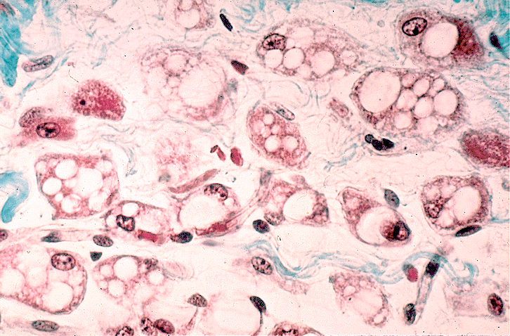
Brown adipocytes contain multiple small lipid droplets (multilocular) and are densely packed with mitochondria with abundant cristae. This is what makes them brown.
Frontiers in Endocrinology comparative anatomy review / 2026
UCP1 (uncoupling protein 1, thermogenin) is located in the inner mitochondrial membrane of brown adipocytes and dissipates the proton electrochemical gradient as heat rather than driving ATP synthesis.
Nicholls DG / FEBS Letters 62:223-228 / 1976
The 1976 Nicholls paper described purine nucleotide control of inner membrane ion conductance in hamster brown adipose tissue, identifying the molecular basis of nonshivering thermogenesis. It is the founding mechanistic paper for UCP1.
Nicholls DG / FEBS Letters / 1976
Cold exposure activates UCP1-mediated thermogenesis via sympathetic innervation of brown adipose tissue. Brown fat is densely innervated; white fat is not.
UCP1 Dependent and Independent Thermogenesis review / Frontiers in Endocrinology / 2020
Long-chain fatty acids both activate UCP1 (by relieving purine nucleotide inhibition) and serve as its substrate.
Mechanism of Fatty-Acid-Dependent UCP1 Uncoupling / PMC / 2013

Beige Adipocyte: The Inducible Type

Beige (brite) adipocytes are a distinct inducible thermogenic cell type that arises within white adipose depots, particularly subcutaneous WAT, in response to cold or beta-adrenergic stimulation. They express UCP1 but are developmentally distinct from classical brown adipocytes.
Wu J, Bostrom P, Sparks LM, Ye L et al. / Cell 150:366-376 / 2012
The Wu 2012 Cell paper identified surface markers TMEM26 and CD137 that distinguish beige adipocytes from classical brown adipocytes in mouse and human tissue. It is the paper that defined the beige cell as a third distinct adipocyte type.
Wu J et al. / Cell / 2012
Beige adipocytes are functionally thermogenic but molecularly distinct from classical interscapular brown adipocytes. Both the terms "beige" and "brite" (brown-in-white) are used; they refer to the same cell.
Harms M and Seale P / Nature Medicine / 2013
Exercise may induce beige adipocyte formation via irisin secreted from muscle.
Bostrom P et al. / Nature / 2012
Adipose browning is increasingly described in disease contexts, not only cold or exercise adaptation. The mechanisms and consequences are still being established.
PubMed 41498391 / 2026
Pharmacologic strategies targeting adipose tissue browning are in active development for obesity and type 2 diabetes.
Frontiers in Pharmacology editorial / 2026

Adipogenesis: The PPARgamma Cascade

PPARgamma is the master transcriptional regulator of adipogenesis. Forced expression in non-adipogenic fibroblasts is sufficient to drive full adipocyte differentiation. This is the most important regulatory fact about how a fat cell becomes a fat cell.
Tontonoz P, Hu E, Spiegelman BM / Cell 79:1147-1156 / 1994
PPARgamma2 is the adipose-specific isoform. Its activation by lipid ligands initiates the full adipogenic transcriptional program.
Tontonoz P, Hu E, Spiegelman BM / Cell / 1994
PPARgamma is required for adipose differentiation both in vitro and in vivo, as demonstrated by knockout studies.
Rosen ED et al. / Molecular Cell / 1999
C/EBPalpha cooperates with and maintains PPARgamma expression; both are required for the full adipogenic program.
Rosen ED et al. / Genes and Development 16:22-26 / 2002
C/EBPbeta and C/EBPdelta are early-response transcription factors that induce C/EBPalpha and PPARgamma at the start of the differentiation cascade.
Farmer SR / Cell Metabolism / 2006

Lipid Droplet Biology: The PLIN Family

The perilipin family (PLIN1-5) are the major structural proteins coating the surface of lipid droplets in adipocytes and other cells.
Bickel PE, Tansey JT, Welte MA / Journal of Lipid Research / 2009
PLIN1 (perilipin-1) is the most abundant lipid droplet coat protein in white adipocytes and the primary PKA substrate during lipolytic stimulation.
Journal of Lipid Research thematic review; Frontiers in Endocrinology / 2011
Under basal conditions, PLIN1 restricts access of cytosolic lipases (HSL, ATGL) to stored triacylglycerol, promoting net lipid storage.
FEBS Letters review on PLIN molecular mechanisms / 2023
Upon catecholamine signaling, PKA phosphorylates PLIN1. CGI-58 is released, activates ATGL (adipose triglyceride lipase), and maximal lipolysis begins.
Multiple PMC reviews
PLIN1 deficiency in mice causes leanness and disordered lipolysis, confirming its essential role in lipid homeostasis.
Tansey JT et al. / Journal of Biological Chemistry (cited in Cardiovascular Research / 2024)

Adipokines

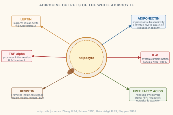
Leptin is a 16-kDa protein encoded by the ob gene, produced primarily by mature adipocytes. Circulating leptin levels reflect adipose mass and act via LEPRb receptors in the hypothalamus to suppress appetite and regulate energy expenditure.
Zhang Y et al. / Nature 372:425-432 / 1994; Annual Reviews leptin review / 2024
Adiponectin (Acrp30) is a 30-kDa C1q-like protein produced exclusively in adipocytes. Its circulating levels are paradoxically reduced in obesity despite increasing adipose mass, and it improves insulin sensitivity.
Scherer PE et al. / Journal of Biological Chemistry / 1995; Berg AH et al. / Nature Medicine / 2001
TNF-alpha is expressed and secreted by adipose tissue and is elevated in multiple obesity models. Neutralization improves insulin-stimulated glucose uptake. This paper established adipose tissue as an inflammatory organ.
Hotamisligil GS, Shargill NS, Spiegelman BM / Science 259:87-91 / 1993
Resistin is an adipocyte-derived hormone that promotes insulin resistance in rodent models. Neutralization of resistin improves glycaemia in obese mice. Its role in human insulin resistance is less established.
Steppan CM et al. / Nature 409:307-312 / 2001

Depot Differences: Visceral vs. Subcutaneous

Visceral adipose tissue (VAT) drains directly to the liver via the portal vein, exposing hepatocytes to elevated free fatty acids from visceral lipolysis and contributing to hepatic insulin resistance. This is the portal hypothesis.
Bjorntorp P / Arteriosclerosis 10:493-496 / 1990
Visceral obesity is an independent cardiometabolic risk factor beyond total adiposity. Bjorntorp named abdominal obesity a "civilization syndrome."
Bjorntorp P / Obesity Research 1:206-222 / 1993
Subcutaneous adipose tissue is metabolically less harmful than visceral. Subcutaneous fat transplantation in mice attenuates metabolic dysregulation associated with visceral fat.
Tran TT et al. / Cell Metabolism / 2008
Depot-specific adipocyte size and adipokine secretion profiles contribute differentially to atherogenic and metabolic risk.
Mancuso P et al. / PMC7815822 / 2021
Adipose depot heterogeneity extends beyond visceral vs. subcutaneous: bone marrow, breast, pericardial, and perivascular depots each carry distinct functions and disease associations.
PMC13467069 / 2026

Histology and Stains

H&E is the most frequently used method for adipose tissue histology, providing general morphological assessment. It was used in 152 of studies in one scoping review.
Histology and Immunohistochemistry scoping review / Cells (MDPI) / 2025 (PMC12190835)
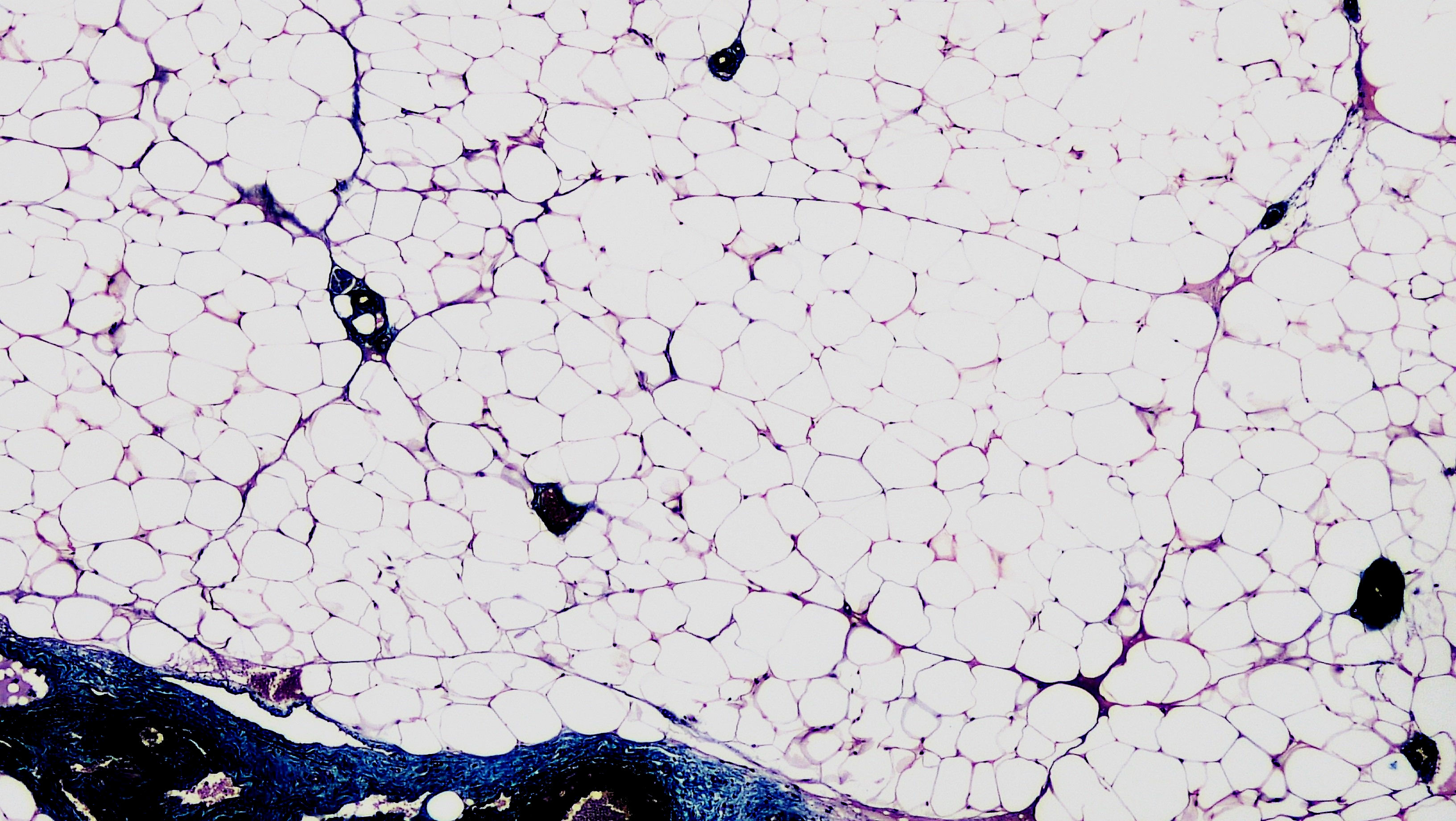
In H&E sections, white adipocytes appear as large empty spaces after lipid extraction by processing solvents. The thin cytoplasmic rim and peripheral nucleus are what survive. The empty space is not pathology; it is the expected artifact of paraffin processing.
Scoping review / Cells / 2025
Oil Red O (ORO) stains neutral lipids red-orange and has largely replaced Sudan III and Sudan IV. It requires frozen sections because paraffin processing removes the lipid ORO would stain.
Scoping review / Cells / 2025
Masson's trichrome stains collagen blue-green and is used to assess fibrosis and stromal composition in adipose tissue.
Scoping review / Cells / 2025
A standardized histological method for quantifying WAT vs. BAT changes under diet challenge was published for mouse models, relevant to anyone trying to define "changed" on a slide with rigor.
PubMed 40983144 / 2025

Processing Failure: This Is the One I Actually Own

I will say this plainly. It is not that fat is fragile out of spite. It is water-heavy, lipid-solvent-sensitive, and structurally almost nothing but membrane around a droplet. Every step in routine processing is built for tissue with more protein scaffolding than fat has. That is not fat's fault. That is a protocol written for a different tissue.

Failure mode Empty-ring artifact, lipid loss · Why it happens Lipid-soluble processing solvents dissolve the droplet content out during dehydration and clearing, leaving a cytoplasmic ring where the cell was.
Leica Biosystems Knowledge Pathway: Processing Fatty Specimens
Failure mode Shrinkage and component loss · Why it happens Paraffin embedding drives out water content, and the same solvents that clear the block strip adipose components beyond just the droplet.
Leica Biosystems Knowledge Pathway: Processing Fatty Specimens
Failure mode Poor sectioning, chatter · Why it happens High lipid content and low protein scaffolding create differential resistance at the blade. Fat lobule borders tear because stroma and fat respond differently to the same sectioning force.
Leica Biosystems Knowledge Pathway: Processing Fatty Specimens
Failure mode Stain mismatch · Why it happens Choice of stain must match the preservation route. ORO requires frozen sections; paraffin has already removed the lipid ORO would stain. Using the wrong stain for the chosen route is a protocol failure, not a tissue failure.
Histology and IHC scoping review / Cells / 2025
Failure mode Milky xylene during clearing · Why it happens Milky or cloudy xylene means water is still present. Dehydration was incomplete. Proceeding to paraffin traps water and guarantees a failed block.
Carson and Hladik / Histotechnology: A Self-Instructional Text, 4th ed.
Failure mode Fat dissolving out is not always failure · Why it happens Surgical pathology labs deliberately dissolve fat with solvents to expose buried lymph nodes. The same mechanism that empties an adipocyte on a slide is used intentionally elsewhere in the same lab. It is proof the solvent works as designed, on schedule or off it.
PMC10927161 / Enzymatic Fat Dissolution

Fix, in short. If lipid preservation is the goal, the paraffin route was already the wrong choice. Freeze the tissue and use a lipid stain. If architecture is the goal, accept that the droplet will dissolve and plan the stain around that instead of fighting it. The block did not fail because fat is difficult. It failed because someone picked a route that was not going to preserve what they needed.

Before labeling something a fat-processing failure, check whether it is a generic processing artifact that happened to land on a fatty block. StatPearls maintains a reference on histology artifacts across tissue types for exactly that differential. StatPearls, Dermatopathology Histology Artifacts


Adipocyte Insulin Signaling

Insulin does not just store glucose. In the adipocyte, it controls the entire balance between lipid storage and release. When this pathway breaks, everything downstream breaks with it.

Insulin binding to its receptor triggers tyrosine autophosphorylation, enabling recruitment and phosphorylation of IRS-1.
White MF / Annual Review of Physiology / 1998
Tyrosine-phosphorylated IRS-1 recruits PI3K (p85 subunit), generating PIP3 at the plasma membrane and activating PDK1.
Vanhaesebroeck B, Alessi DR / Biochemical Journal / 2000
PDK1 phosphorylates Akt (Ser473/Thr308). Activated Akt phosphorylates AS160/TBC1D4, releasing its GAP activity and allowing Rab10-driven GLUT4 vesicle translocation to the plasma membrane in adipocytes.
Sano H et al. / Journal of Cell Biology / 2003
Adipocytes rely almost exclusively on the canonical PI3K-Akt-TBC1D4-Rab10 axis for GLUT4 translocation, unlike muscle cells that also have an AMPK-dependent exercise route.
Fazakerley DJ et al. / Cell Metabolism / 2025 (PMC13116035)
Akt2 is the dominant isoform mediating insulin-stimulated GLUT4 translocation in adipocytes. Akt2-null mice are insulin resistant.
Cho H et al. / Science / 2001
Insulin also inhibits lipolysis via Akt-mediated phosphorylation of PDE3B, raising cAMP hydrolysis, lowering PKA activity, and inactivating HSL.
Degerman E et al. / Journal of Biological Chemistry / 1998

Adipocyte Insulin Resistance: What Breaks Down in Obesity

The fat cell does not become insulin resistant because it is lazy. It becomes insulin resistant because it is inflamed, overwhelmed, and serine-phosphorylated into a state where the signal cannot get through.

TNF-alpha from adipose macrophages activates JNK1, which serine-phosphorylates IRS-1 (Ser307 in mice, Ser312 in humans), blocking its interaction with the insulin receptor. This is the molecular link between adipose inflammation and insulin resistance.
Hotamisligil GS, Shargill NS, Spiegelman BM / Science 259:87-91 / 1993
Serine phosphorylations in the IRS-1 PIR domain directly abrogate IRS-1 and insulin receptor interaction.
Ye Z et al. / PNAS / 2024
IKKbeta/NF-kB pathway activation by saturated fatty acids and TNF-alpha independently induces IRS-1 serine phosphorylation in adipocytes.
Arkan MC et al. / Nature Medicine / 2005
ER stress in hypertrophic adipocytes activates IRE1alpha and PERK, which phosphorylate JNK and IKKbeta, converging on IRS-1 serine phosphorylation.
Ozcan U et al. / Science / 2004
Ceramides, generated from excess saturated fatty acid flux, directly inhibit Akt by activating PP2A and recruiting Akt to PKCzeta scaffolds, blocking Akt membrane translocation.
Summers SA / Progress in Lipid Research / 2006
Chronic adipose inflammation with macrophage-derived IL-6 upregulates SOCS3, which targets IRS-1 for ubiquitin-mediated proteasomal degradation.
Lebrun P, Van Obberghen E / Biochemical Journal / 2008
Insulin-stimulated glucose uptake in adipocytes is blunted by 60 to 80% in obesity, tracking with reduced IRS-1 tyrosine phosphorylation and PI3K activity.
Rondinone CM et al. / Diabetes / 1997

Lipolysis Regulation in Disease

ATGL (PNPLA2) is the rate-limiting triglyceride hydrolase in adipocytes, performing the first step of triacylglycerol hydrolysis to diacylglycerol and fatty acids. ATGL-null mice show massive lipid accumulation.
Zimmermann R et al. / Science 306:1383 / 2004
HSL performs the second step (DAG to MAG) and is the primary catecholamine-activated lipase. PKA phosphorylates HSL (Ser563, Ser660) and PLIN1, allowing lipase access to the lipid droplet.
Holm C / Biochemical Society Transactions / 2003
PLIN1 phosphorylation by PKA releases CGI-58 (co-activator of ATGL), coordinately activating both ATGL and HSL upon adrenergic stimulation.
Granneman JG et al. / Journal of Biological Chemistry / 2009
Insulin suppresses lipolysis by activating PDE3B via Akt, lowering cAMP, reducing PKA activity, and re-esterifying PLIN1. This anti-lipolytic action is severely impaired in obese adipocytes.
Degerman E et al. / Journal of Biological Chemistry / 1998
Basal (non-stimulated) lipolysis is elevated in obesity, correlated with adipocyte size and local inflammation. Elevated fasting FFA flux is a key driver of hepatic and peripheral insulin resistance.
Langin D et al. / International Journal of Obesity / 2006

Lipotoxicity: When the Fat Cell Overflows

The term "lipotoxicity" was coined by Roger Unger to describe how lipid overload of pancreatic islets causes beta-cell dysfunction in Zucker diabetic fatty rats.
Unger RH / Diabetes 44:863 / 1995
When adipose tissue reaches its safe storage limit (the adipose expandability threshold), excess lipid overflows to liver, skeletal muscle, heart, and pancreas, causing organ-specific insulin resistance and injury.
Virtue S, Vidal-Puig A / Cell Metabolism / 2010
Intramyocellular lipid accumulation, particularly diacylglycerol and ceramide species, activates PKC-theta in muscle, impairing IRS-1 signaling and reducing insulin-stimulated glucose uptake.
Itani SI et al. / Diabetes / 2002
Hepatic lipid accumulation activates PKC-epsilon, impairing insulin receptor kinase signaling and increasing hepatic glucose output. This is a key mechanism linking adipose overflow to T2D.
Samuel VT et al. / Journal of Clinical Investigation / 2007
Pancreatic beta-cell lipotoxicity is glucose-dependent (glucolipotoxicity): high glucose combined with elevated FFAs synergistically impairs insulin secretion and promotes apoptosis.
Prentki M et al. / Diabetes / 2001

Adipose Inflammation in Obesity

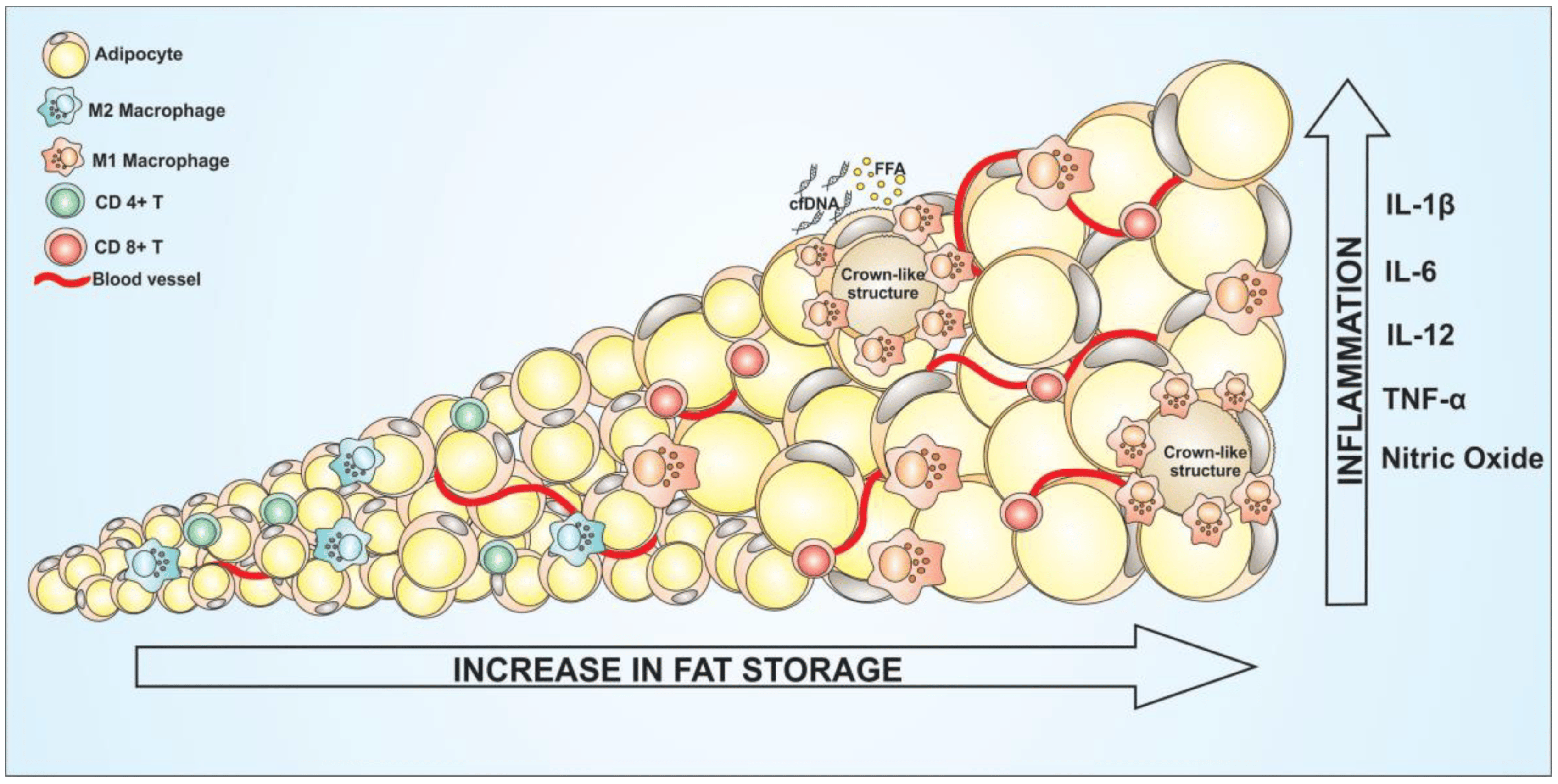
The macrophage is not an innocent bystander in fat. In obesity it is an active participant, and the crown it forms around a dying adipocyte is visible on a slide.

Macrophages constitute up to 40% of all cells in obese adipose tissue. Adipose macrophages account for nearly all TNF-alpha and significant IL-6 expression in fat. Macrophage number correlates with adipocyte size and BMI.
Weisberg SP, McCann D, Desai M et al. / Journal of Clinical Investigation 112:1796 / 2003
Obesity in mice and humans is associated with chronic inflammation in fat, driven by upregulation of macrophage-specific and inflammation genes in white adipose tissue. Proposed as a cause of obesity-related insulin resistance.
Xu H, Barnes GT, Yang Q et al. / Journal of Clinical Investigation 112:1821 / 2003
Crown-like structures (CLS) form when macrophages surround and engulf dead adipocytes in obese fat. CLS density correlates with systemic insulin resistance and hyperinsulinemia in humans.
Cinti S et al. / Journal of Lipid Research / 2005
Obesity induces a phenotypic switch in adipose macrophages from anti-inflammatory M2 (CD163+, IL-10-secreting) toward pro-inflammatory M1 (CD11c+, TNF-alpha/IL-6-secreting).
Lumeng CN, Bodzin JL, Saltiel AR / Journal of Clinical Investigation / 2007
The NLRP3-Caspase-1 inflammasome is active within CLS in human adipose tissue and contributes to IL-1beta production and insulin resistance.
PMC12877315 / Obesity Surgery / 2025

Adipocyte Hypertrophy vs. Hyperplasia

Adipocyte size is inversely correlated with insulin sensitivity independent of total adiposity. Smaller adipocytes show greater insulin-stimulated glucose uptake and lower basal lipolysis.
Weyer C et al. / Journal of Clinical Investigation / 2000
Enlarged adipocytes secrete elevated IL-6, IL-8, MCP-1, and leptin while producing less adiponectin. The inflammatory secretome is a direct function of cell size, not just depot mass.
Skurk T et al. / Journal of Clinical Endocrinology and Metabolism / 2007
Hypertrophic adipose expansion is associated with inflammation, ER stress, and insulin resistance even after adjusting for body composition.
Alligier M et al. / International Journal of Molecular Sciences / 2023
Hyperplastic adipose expansion (more smaller cells) is metabolically favorable compared to hypertrophic expansion (fewer larger cells). Pharmacological promotion of hyperplasia is a proposed therapeutic strategy.
Tchoukalova YD et al. / Diabetes / 2010
The distinction between healthy (hyperplastic) and unhealthy (hypertrophic) obesity explains why some obese individuals remain metabolically normal.
McLaughlin T et al. / Journal of Clinical Endocrinology and Metabolism / 2007

Adiponectin Deficiency and Insulin Resistance

Plasma adiponectin is inversely correlated with obesity, visceral fat mass, and insulin resistance. Levels are reduced by 50 to 70% in obese and T2D individuals despite increasing adipose mass.
Arita Y et al. / Biochemical and Biophysical Research Communications / 1999
Adiponectin activates AMPK in skeletal muscle via AdipoR1, increasing fatty acid oxidation and glucose uptake, and activates PPARalpha in liver via AdipoR2, reducing gluconeogenesis.
Yamauchi T et al. / Nature Medicine / 2002
AdipoR1/AdipoR2 double-knockout mice develop insulin resistance, inflammation, and oxidative stress identical to adiponectin-null mice, confirming receptor specificity.
Yamauchi T et al. / Nature Medicine / 2007
Adiponectin suppresses NF-kB activation in macrophages and endothelial cells, linking its deficiency to both insulin resistance and cardiovascular risk.
Ouchi N et al. / Circulation / 2000
Thiazolidinediones increase adiponectin secretion from adipocytes via PPARgamma activation, and this is considered a primary mechanism of their insulin-sensitizing effect.
Maeda N et al. / Nature Medicine / 2001

Adipocyte Dysfunction in Type 2 Diabetes

Enlarged diabetic adipocytes are resistant to insulin's antilipolytic effect, producing day-long elevated plasma FFAs that stimulate hepatic gluconeogenesis and peripheral insulin resistance.
Boden G / Metabolism / 1997
White adipose tissue in T2D shows downregulated GLUT4 gene expression, one of the earliest molecular defects detected, accounting for reduced insulin-stimulated glucose disposal.
Shepherd PR, Kahn BB / New England Journal of Medicine / 1999
Dysfunctional T2D adipocytes overproduce leptin, TNF-alpha, resistin, and IL-6 while underproducing adiponectin. This adipokine imbalance amplifies systemic insulin resistance.
Rabe K et al. / Diabetes Care / 2008
Impaired de novo lipogenesis and reduced lipid re-esterification in T2D adipocytes force lipid overflow to non-adipose tissues.
DeFronzo RA et al. / Journal of Clinical Endocrinology and Metabolism / 2004
A 2025 review identifies four core defects in T2D adipocytes: impaired insulin signaling, altered adipokine secretion, mitochondrial dysfunction, and enhanced basal lipolysis.
Rohm TV et al. / Diabetology and Metabolic Syndrome / 2025

Metabolic Syndrome and Adipose

Visceral adipose tissue, not subcutaneous adipose tissue, drives the metabolic syndrome cluster. VAT volume independently predicts insulin resistance, dyslipidemia, and T2D risk after adjusting for BMI.
Despres JP, Lemieux I / Nature / 2006
VAT adipocytes are more lipolytically active, less responsive to insulin's antilipolytic action, and drain FFAs directly into the portal circulation. This gives portal FFA flux a primary role in hepatic insulin resistance.
Jensen MD / Diabetes Care / 2006
Adipose tissue insulin resistance is a critical early event that initiates metabolic syndrome, not merely a consequence of it.
PMC12561392 / Frontiers in Endocrinology / 2025
Metabolic syndrome is best understood as a disease of adipose tissue dysfunction rather than simply excess fat mass. Individuals with metabolically healthy obesity retain functional adipose tissue capable of lipid buffering.
Eckel RH et al. / Lancet / 2010

Recent Literature 2024 to 2026

Single-nucleus RNA sequencing of subcutaneous and visceral adipose depots identified mesothelial cells, adipocytes, and adipocyte-progenitor cells as the cell types most strongly correlated with metabolic disease severity.
Becker M et al. / Cell Metabolism / 2024
snRNA-seq identified at least two distinct beige adipocyte subpopulations: UCP1-beige and FC (futile-cycle) adipocytes, with distinct thermogenic and metabolic gene programs.
Jang C et al. / Cell Metabolism / 2024
An integrative snRNA-seq analysis of subcutaneous adipose from 84 individuals with metabolic syndrome identified WNT signaling from progenitor cells to mature adipocytes as a key differentiation regulator.
PMC12542196 / Diabetology and Metabolic Syndrome / 2025
Depot-specific adipose traits (cellular composition, inflammatory status, developmental origin) outperform BMI alone in predicting cardiometabolic risk across multiple diseases.
PMC13100352 / Frontiers in Cell and Developmental Biology / 2026
GLP-1 receptor agonist resistance in obesity management is a newly recognized clinical challenge. A significant subset of patients exhibits no weight loss or weight regain, suggesting adipocyte-intrinsic resistance mechanisms not yet characterized.
PMC13236842 / 2025
snRNA-seq of human WAT identifies a low-maturation adipocyte subpopulation defined by TSHZ3-mediated transcriptional suppression; this subset is enriched in metabolic disease and represents a new axis of intra-depot heterogeneity beyond depot location.
Efthymiou V et al. / Nature Communications / 2026 (PubMed)
NPY receptor 1 (NPY1R) was identified as a cell-autonomous brake on adipocyte lipolysis by single-cell transcriptomics of human weight loss; NPY1R expression inversely correlates with BMI and rises after weight loss, linking neuropeptide Y signaling directly to lipolysis control in the fat cell.
Grothen JER et al. / Molecular Metabolism / 2026 (PubMed)
Defective subcutaneous adipogenesis drives insulin resistance in non-obese individuals, demonstrating that adipocyte dysfunction, not adipose mass, is the primary adipose-origin cause of metabolic disease.
Frontiers in Physiology / 2025
A peroxisomal pathway involving monomethyl branched-chain fatty acid metabolism drives thermogenesis in fat cells independently of UCP1, opening a new target class for browning strategies.
Nature Reviews Endocrinology / 2025; nature.com/articles/s41574-025-01193-x
Leptolin was identified as a novel adipocyte-derived adipokine positively correlated with exercise and inversely with BMI; it increases energy expenditure in animal models and may extend the canonical adipokine panel.
PMC13335978 / 2026
UCP1-IRES-Cre knock-in mice enable selective brown adipocyte targeting without CNS off-target expression; prior Cre lines had neuronal activity that confounded thermogenesis phenotyping.
PMC13308379 / 2026
Adipogenin's role in seipin-mediated lipid droplet enlargement is now framed as a new paradigm: lipid droplet architecture itself, not just lipid content, contributes to metabolic disease risk and is emerging as a cancer link.
PubMed 42005040 / 2026
FPLD2 (familial partial lipodystrophy type 2) snRNA-seq reveals suppressed lipid and mitochondrial gene programs alongside elevated inflammation in remaining adipocytes; the adipocyte is failing before it disappears from the depot.
PMC12721891 / 2026
Adipose tissue macrophage heterogeneity is more complex than M1/M2 polarization: single-cell data reveal multiple activation states with distinct metabolic roles in obesity-driven inflammation.
PMC12937715 / 2026
Three druggable targets at the fat cell -- AMPK, PKM2, and UCP1 -- are now synthesized in a single pharmacology review; the first systematic ranking of direct adipocyte drug targets by mechanism class.
Yang et al. / Biochemical Pharmacology / 2026 (PubMed)
Ferroptosis -- iron-dependent regulated cell death -- in adipocytes is proposed as a systemic metabolic regulator, not just a local cell-death pathway; disrupted iron handling in fat may drive systemic metabolic consequences.
Wang et al. / Science Bulletin / 2026 (PubMed)
Autophagy suppression during thermogenic activation promotes mitochondrial accumulation in brown and beige adipocytes; enhanced autophagy contributes to thermogenic decline, making autophagy flux a direct regulator of fat cell heat output.
Villarroya et al. / Int Rev Cell Mol Biol / 2026 (PubMed)

Classic Papers

Paper Leptin / ob gene cloning · Authors Zhang Y, Proenca R, Maffei M, Barone M, Leopold L, Friedman JM · Journal / Year Nature 372:425-432 / 1994 · What It Established Leptin is a 16-kDa adipocyte-secreted hormone regulating energy balance. Established adipose as endocrine.
Paper PPARgamma as master adipogenic regulator · Authors Tontonoz P, Hu E, Spiegelman BM · Journal / Year Cell 79:1147-1156 / 1994 · What It Established PPARgamma2 expression in fibroblasts drives full adipocyte differentiation. Master regulator defined.
Paper UCP1 uncoupling mechanism · Authors Nicholls DG · Journal / Year FEBS Letters 62:223-228 / 1976 · What It Established Purine nucleotide control of inner membrane conductance in brown fat mitochondria. Founding UCP1 paper.
Paper Acrp30 / adiponectin discovery · Authors Scherer PE, Williams S, Fogliano M, Baldini G, Lodish HF · Journal / Year Journal of Biological Chemistry 270:26746-26749 / 1995 · What It Established Novel 30-kDa C1q-like adipocyte protein, induced 100-fold during differentiation. Second major adipokine.
Paper TNF-alpha from adipose tissue · Authors Hotamisligil GS, Shargill NS, Spiegelman BM · Journal / Year Science 259:87-91 / 1993 · What It Established Adipose tissue expresses and secretes TNF-alpha in obesity. Established fat as inflammatory.
Paper Beige adipocyte identification · Authors Wu J et al. (Spiegelman lab) · Journal / Year Cell 150:366-376 / 2012 · What It Established Beige adipocytes are a distinct thermogenic cell type in WAT, identified by TMEM26/CD137 surface markers.
Paper Resistin links obesity to diabetes · Authors Steppan CM et al. · Journal / Year Nature 409:307-312 / 2001 · What It Established Adipocyte-secreted resistin promotes insulin resistance; neutralization improves glycaemia in obese mice.
Paper Portal / visceral fat hypothesis · Authors Bjorntorp P · Journal / Year Arteriosclerosis 10:493-496 / 1990 · What It Established Visceral fat drains portal FFA to liver, driving hepatic insulin resistance. Canonical depot-risk paper.

Scan log

Date PMIDs added Notes
2026-09-13 42728326, 42726295, 42721855, 42721252, 42716945 9 PMIDs returned for last 72 h; 5 relevant to adipocyte biology added; 4 excluded (nephropathy, hepatocyte exosomes, breast cancer spatial multi-omics, all off-target)

Latest 8

2026-09-10 · Nature Communications
Single-nucleus analysis of human white adipose tissue reveals adipocyte subsets with distinct metabolic profiles

Efthymiou V et al. 2026. snRNA-seq of human WAT identifies adipocyte subpopulations; a low-maturation subset linked to TSHZ3-mediated transcriptional suppression is associated with metabolic disease severity.

2026-09-10 · Biochemical Society Transactions
Understanding adipocyte heterogeneity across species, depot, and disease

Kitto ES et al. 2026. snRNA-seq and spatial transcriptomics reveal previously unappreciated adipocyte subpopulations across depots and disease states; cross-species comparisons clarify conserved vs. human-specific subsets.

2026-09-10 · Molecular Metabolism
Single cell transcriptomics of human weight loss links adipocyte NPY1R to control of lipolysis

Grothen JER et al. 2026. NPY receptor 1 (NPY1R) identified as a cell-autonomous brake on adipocyte lipolysis; expression is inversely correlated with BMI and rises after weight loss, implicating neuropeptide Y signaling in direct lipolysis control.

2026-09-10 · Physiology
Adipose Progenitor Cells in Thermogenesis and Metabolic Regulation

Sahin C et al. 2026. Heterogeneous adipose progenitor subsets are characterized; specific subpopulations are required for thermogenic adipocyte generation and healthy remodeling, with implications for browning strategies.

2026-09-10 · Physiological Reports
Adipocyte-specific FFA2 deletion leads to increased adipose inflammation and altered intestinal lipid handling

Nnyamah C et al. 2026. Free Fatty Acid Receptor 2 (FFA2/FFAR2) on adipocytes suppresses adipose tissue inflammation; mice lacking adipocyte FFA2 show worsened inflammatory phenotype and disrupted intestinal lipid absorption.

2026-09-10 · Frontiers in Physiology
Impaired adipogenesis drives insulin resistance in non-obese individuals

Defective subcutaneous adipogenesis produces insulin resistance independently of excess adipose mass, expanding the adipocyte dysfunction framework beyond obesity to lean insulin-resistant individuals.

2026-09-10 · PMC
Adipocyte size, overweight, and insulin resistance in T2DM: systematic review

Systematic review of 113 human and 29 animal studies: adipocyte hypertrophy is consistently associated with insulin resistance independent of total adiposity; weight loss reduces cell size and improves insulin sensitivity in parallel.

2026-09-10 · PMC
Adipocyte browning: mechanisms and anti-obesity pharmacology

Reviews white-to-beige adipocyte transdifferentiation mechanisms and current pharmacological strategies including beta-3 agonists, thyroid hormone analogs, and FGF21 mimetics targeting browning for obesity treatment.